Most of this information is in addition to what is listed on our web-site.
Radiation (Oncology) Masks and Burn Masks
There are two primary treatments for which a hard plastic mask is fabricated for a medical
patient. The first is for radiation treatments. There are two purposes, one is to define the region
in which the radiation is to be directed (though a hole in the mask), the other is too provide an
energy “filter” so that radiation of the proper energy impacts the area of interest, the skin for
example in skin cancer patients.
The other application is for burn masks. These plastic masks are used in the treatment of burn
patients to minimize the disfigurement due to scare tissue. Patients wear these masks, usually
with special lotions underneath, for up to 23 hours per day for many months.
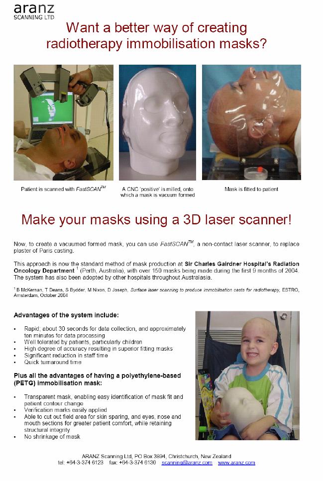
Most of these masks are fabricated today using a very old and messy process of applying wet
plaster to the face, waiting for it to dry and then removing it. This forms a cast from which the
plastic mask can be made. The current wet plaster method is very slow and very uncomfortable
for the patient. See the following description of this process:
http://www.regionshospital.com/Regions/Menu/0,,11383,00.html
The FastSCAN is excellent for this application because it is very fast, accurate and very
comfortable for the patient. The comfort factor is particularly important with children.
Please see the following document concerning the use of the FastSCAN for making masks:
(note: the FastSCAN for Oncology software is not required for this application. It only provides
some shortcut keys for some of the functions. Also, this is not currently offered by Polhemus.
We are in discussions with Aranz about this.)
Orthotics and Prosthetics
Of course the best known example of this application is from our customer Hanger
prosthetics in the world. In a way similar to the fabrication of face masks, the traditional method
of fabricating an artificial limb involves making a replica of the residual limb using the plaster
cast method. Using the replica residual limb, an artificial extension (prosthetic) can be fitted.
By using the FastSCAN, the plaster cast process can be eliminated, resulting in faster turnaround
of prosthetics for the patient, as well as a more comfortable fitting process for the patient. In
addition, having a digital record of the patient’s residual limb eliminates the need for care and
storage of a plaster cast model.
There is also a growing interest in using the FastSCAN for foot orthotics which are inserts for
the shoe to correct for foot abnormalities or improper alignment of the foot. Currently the shoe
inserts are made in a similar fashion as the face masks by first making a wet plaster cast. From
this plaster cast shoe inserts are fabricated. Again, the FastSCAN is faster, more accurate and
more comfortable for the patient.
Cosmetic and Plastic Surgery
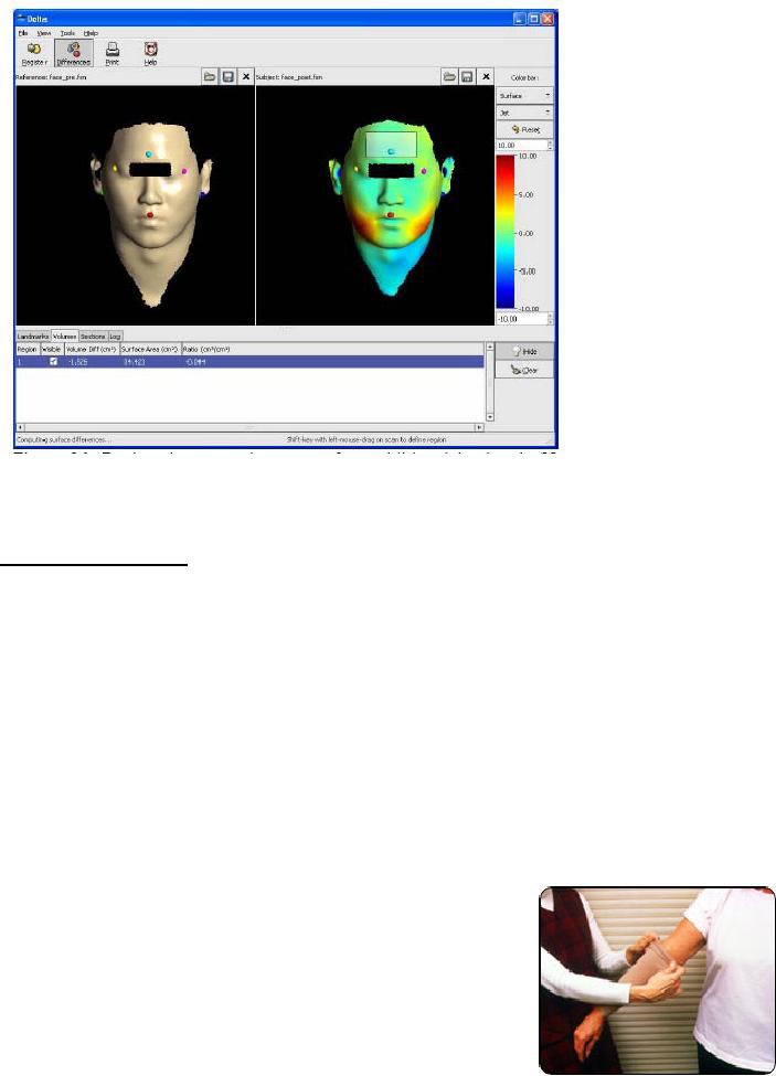
The use of 3D modeling in cosmetic and plastic surgery is growing very rapidly. In both cases it
is very useful to have a 3D image of the patient before and after the surgery. For example, if a
person wants to improve their appearance by having an injection of filler to reduce skin wrinkles
(with Botox for example), the FastSCAN images could be used to measure the result by using
the DeltaView software. A theoretical example is shown below of before and after images which
were imported into DeltaView to measure the volume change. (Note the swelling in the cheeks):
This allows the doctors and surgeons to quantify the results of their procedures and to provide
verification to the patients that a change did occur.
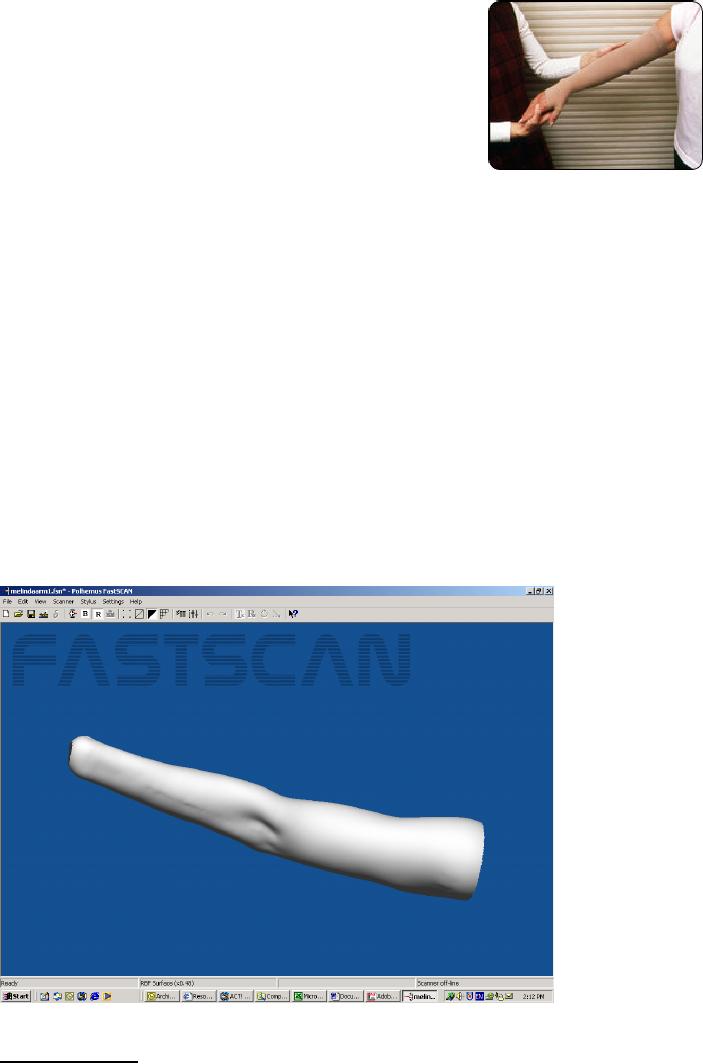
Compression Garments
Compression garments are tight fitting pieces of special clothing used to help reduce swelling
(particularly in arms and legs) that can occur in patients with lymphoedema or as a result of
surgery. Currently the measurements for these garments are done manually with a flexible tape
measure. This method is slow and prone to errors. By using the FastSCAN, accurate, fast and
highly repeatable measurements can be made. The scan files can then be e-mailed to the
manufacturer to cut the cloth and make the garment. Please see the following excerpt from a
Note in the first picture below the use of a flexible tape measure for measuring the arm. Many
measurements would be taken along the arm to create a custom garment. The pressure applied to
the tape and the exact location of the measurements would vary from technician to technician
and are sometime made with errors.
Many patients will be prescribed garments that will provide
compression for the affected limb. The garments help to keep
fluid from accumulating in the limb. These garments have
specific amounts of pressure and can be worn on the legs,
hands, feet, or arms. The garments are made of a tight
stretchy fabric. An expert fitter must fit lymphedema
garments (sleeves). Measurements are taken, and a patient
must try on the sleeves to make certain that they have a
comfortable fit. Sometimes custom sleeves must be made, but
most people are able to find a pre-made sleeve in a suitable
size. The sleeves prevent the accumulation of more fluid in the limb; they do not pump fluid
out of the limb. The garments are usually used in combination with therapy or as a
preventive or maintenance measure.
Lymphedema sleeves and treatment can change the size of the affected limb as can various
activities. Sometimes patients need more than one sleeve during this process because of
the changing size of the limb. There is a tendency for patients to think that their sleeve has
been fit improperly. Sometimes it has been, but more often than not, the limb has changed
in size.
Note these sleeves wear out with continued daily use and must be refit and replaced on a
regular basis (approximately every 3-6 months). Over time with washing and wearing they
lose their compression. Different levels of compression are used for prevention versus
maintenance.
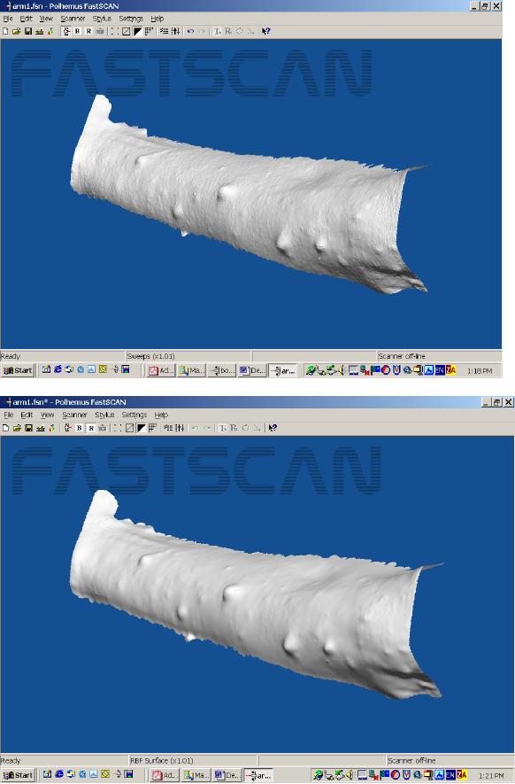
Below is a FastSCAN image for use in creating a compression garment for the arm:
Other Applications
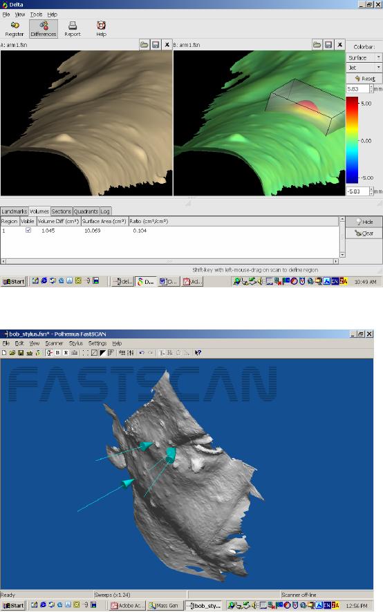
There are many more applications for the FastSCAN which come from individual doctors which
may or may not become widespread applications. A recent example comes from a neurologist
who wants to map the location of neurofibrosis tumors (see images below) and track their
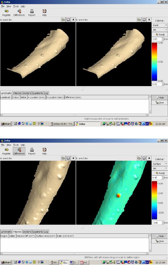
reduction in size as a result of his treatment. For this he intends to use the FastSCAN with a
stylus to map the tumor locations and DeltaView to measure the change in size/volume.
Raw Scan – right arm.
RBF processed image – right arm.
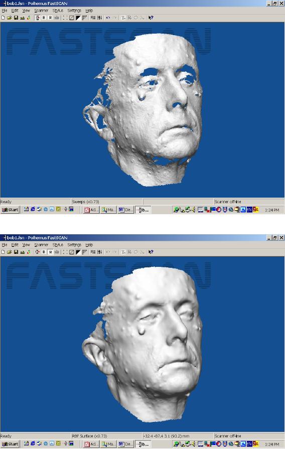
Raw Scan.
RBF Process Image.
DeltaView software – baseline images loaded.
DeltaView – color topography.
DeltaView – Cross section calculation showing tumor diameter measurement.
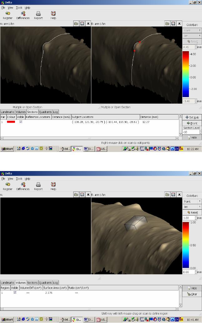
DeltaView – Surface area calculation.
DeltaView – Volume calculation.
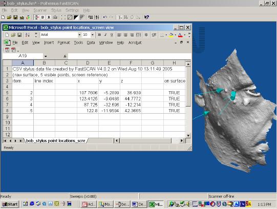
FastSCAN – Stylus points.
FastSCAN – Stylus points export file (Excel format) showing x,y,z locations of each stylus point.